THIS PRODUCT IS DISCONTINUED
Fast Activated Cell-based ELISA (FACE™) Kits provide a simple, sensitive method for detecting protein phosphorylation directly in the cell, without making extracts or performing electrophoresis and membrane blotting. These 96-well, high-throughput assays are available in both colorimetric and chemiluminescent formats for over 20 different targets (see list at right). For complete details, click the FACE™ Method tab below.
FACE PI3 Kinase p85 Kits provide 96 rxns each of 2 antibodies that enable you to monitor and compare the levels of both phosphorylated and total PI3K p85. The phospho-PI3K p85 antibody preferentially recognizes peptides and proteins containing phospho-tyrosine at the consensus YXXM motif. The total-PI3K p85 antibody recognizes PI3K p85 regardless of its phosphorylation state. Click the PI3K p85 Info tab below for data and more information.
| Name | Format | Cat No. | Price | |
|---|---|---|---|---|
| FACE™ PI3 Kinase p85 | 1 x 96 rxns | 48175 | Discontinued | |
| 5 x 96 rxns | 48675 | Discontinued | ||
| FACE™ PI3 Kinase p85 Chemi | 1 x 96 rxns | 48275 | Discontinued | |
| 5 x 96 rxns | 48775 | Discontinued | ||
| FACE™ PI3 Kinase p85 Manual |
| FACE™ Profile |
| Cell Biology Products Brochure |
| IsoCyte™ Application Note – Phospho-Protein Detection |
| MSDS: Sodium Azide |
| MSDS: Sulphuric Acid |
| MSDS: Thimersol |
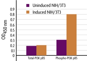
Figure 1: Measurement of phosphorylated and total PI3 Kinase.
NIH/3T3 cells were cultured in 96-well plates and serum-starved for 16 hours. Cells were then treated with 50 ng/ml of PDGF for 5 minutes and fixed. Total and phospho PI3K p85 were each assayed in triplicate using the phospho and total PI3K p85 antibodies included in the FACE PI3 Kinase Kit. Data was plotted after correction for cell number (performed through use of Crystal Violet).
Antibody Specificities
The phospho-PI3K p85 antibody preferentially recognizes peptides and proteins containing phospho-tyrosine at the consensus YXXM motif. It was raised against a synthetic phospho-(Tyr) p85 binding motif peptide. Some cross-reactivity is observed for peptides that contain phospho-tyrosine followed by methionine. No cross-reactivity is observed with corresponding nonphosphorylated sequences or with other phospho-tyrosine-containing motifs. The total-PI3K p85 antibody recognizes the total level of PI3K p85 proteins regardless of the phosphorylation state.
PI3 Kinase p85 Overview
Phosphoinositide 3 Kinase (PI3 Kinase, PI3K) is a regulator of cell proliferation, survival and growth. PI3 Kinase catalyzes the production of phosphatidylinositol-3,4,5-triphosphate by phosphorylating phosphatidylinositol (PI), phosphatidylinositol-4-phosphate (PIP) and phosphatidylinositol-4,5-biphosphate (PIP2). Growth factors and hormones are responsible for triggering this phosphorylation event and PH domain-containing signaling molecules, including various kinases and their regulators, recognize the resulting product. This activates corresponding cell signaling pathways, eventually leading to cell growth, cell cycle entry, cell migration and cell survival.
The FACE™ Method
In FACE, cells are cultured in 96-well plates and stimulated to induce the pathway of interest. Following stimulation, the cells are fixed rapidly, which preserves activation-specific protein modifications. Each well is then incubated with a primary antibody specific for the activated protein of interest. Subsequent incubation with secondary HRP-conjugated antibody and developing solution provides a colorimetric or chemiluminescent readout that is quantitative and reproducible (Figure 1). The number of cells in each well can be normalized easily with the provided Crystal Violet solution. FACE Kits also contain primary antibody specific for the native inactive protein, so you can monitor both native and activated protein levels in the same experiment. FACE eliminates cellular extractions, radioactive kinase assays, time-consuming Westerns and inefficient epitope interactions that occur on membranes. FACE is a highly sensitive high-throughput assay designed for detecting activated proteins within mammalian cells.
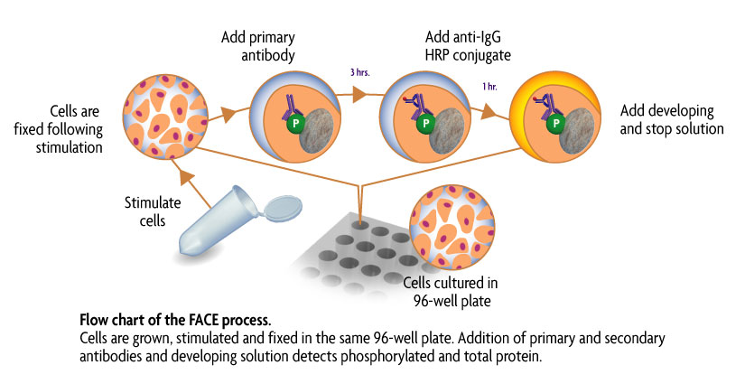
Figure 1: Flow chart of the FACE process.
Flow chart of the FACE in cell Western method that uses a cell based ELISA to measure the levels of the native and phospho forms of signaling proteins and kinases that are activated by phosphorylation.
Contents & Storage
Two (or ten) 96-well plates for culturing cells, 96 (or 5 x 96) rxns each of two primary antibodies (1 phospho-specific, 1 specific for native protein), HRP-conjugated secondary antibody, Quenching Solution, 1X Antibody Blocking Buffer, 1X Antibody Dilution Buffer, 10X PBS, 10% Triton X-100, 1% SDS Solution, Developing and Stop Solutions, and Crystal Violet Cell Quantification Solution. Storage conditions vary from room temperature to -20°C, see manual for details. All reagents are guaranteed stable for 6 months when stored properly.