<< Back to Targets & Applications Home Page
Histone Modifications Guide
Information regarding histone modifications, their biological relevance and the proteins involved in their regulation.
The three billion base pairs of DNA in each of your cells are incorporated into chromatin by associating with a group of small evolutionarily conserved proteins, the histones. Two molecules each of the core histone proteins (H2A, H2B, H3 and H4) form an octamer where ~147 bp of DNA wraps around it to create the fundamental repeating subunit of chromatin, the nucleosome. A specialized histone (linker histone H1) functions to further compact the tens of millions of nucleosomes into more highly ordered structures. It is this compacted DNA-protein complex that serves as the template for all DNA-dependent processes including DNA replication, repair, recombination, chromosome segregation and gene expression.
It has long been known that the histones are subject to a variety of post-translational modifications (phosphorylation, acetylation, ubiquitylation, methylation: two different types – arginine and lysine) and that these modifications play important roles in chromatin structure and function. With regard to transcriptional regulation, increases in histone acetylation and specific methylation events (H3 lysine 4 & lysine 36 methylation) are generally associated with increased gene expression, while decreased acetylation and other histone methylation events (H3 lysine 9 and lysine 27 methylation) are hallmarks of decreased gene expression (See reference 6 for a recent review).
Histone Modifications Products
-
Histone and histone modification antibodies
-
Histone modifying enzymes
-
Histone modification ELISAs
-
Histone purification kits
Histone Modifications Map
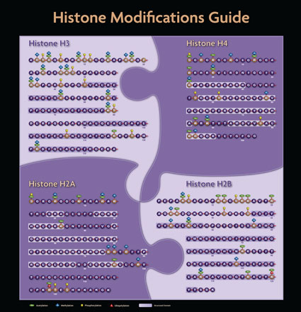
Click here to download the Histone Modifications Guide.
Histone Modifications and Associated Biological Function
| Site | Modification | Associated Function |
| Ser1 | Phosphorylation | Mitosis |
| Lys5 | Acetylation | Transcriptional activation |
| Lys119 | Ubiquitylation | Spermatogenesis |
| Thr120 | Phosphorylation | Mitosis |
| Site | Modification | Associated Function |
| Ser139 | Phosphorylation | DNA Damage repair, apoptosis |
| Tyr142 | Phosphorylation | Regulation of DNA damage foci formation |
| Site | Modification | Associated Function |
| Lys5 | Acetylation | Transcriptional activation |
| Lys12 | Acetylation | Transcriptional activation |
| Ser14 | Phosphorylation | Apoptosis |
| Lys15 | Acetylation | Transcriptional activation |
| Lys20 | Acetylation | Transcriptional activation |
| Lys120 | Ubiquitylation | Transcription (elongation?) |
| Site | Modification | Associated Function |
| Arg2 | Methylation | Transcriptional activation |
| Thr3 | Phosphorylation | Mitosis |
| Lys4 | Acetylation | Transcriptional activation |
| Lys4 | Methylation | Euchromatin, transcriptional activation |
| Thr6 | Phosphorylation | Transcriptional activation |
| Arg8 | Methylation | Transcriptional activation |
| Lys9 | Acetylation | Histone deposition, Transcriptional activation |
| Lys9 | Methylation | Transcriptional silencing, heterochromatin |
| Ser10 | Phosphorylation | Mitosis, immediate early gene activation |
| Thr11 | Phosphorylation | Mitosis; DNA damage induced transcription |
| Lys14 | Acetylation | Transcriptional activation |
| Arg17 | Methylation | Transcriptional activation |
| Lys18 | Acetylation | Transcriptional activation |
| Lys23 | Acetylation | Transcriptional activation |
| Arg26 | Methylation | Transcriptional activation |
| Lys27 | Methylation | Transcriptional silencing |
| Ser28 | Phosphorylation | Mitosis |
| Lys36 | Acetylation | Transcription activation |
| Lys36 | Methylation | Transcription elongation |
| Thr45 | Phosphorylation | DNA replication, apoptosis |
| Lys56 | Acetylation | DNA damage repair, chromatin assembly |
| Lys79 | Methylation | Transcriptional activation |
| Site | Modification | Associated Function |
| Ser1 | Phosphorylation | Transcriptional activation |
| Arg3 | Methylation | Transcriptional activation |
| Lys5 | Acetylation | Histone deposition, Transcriptional activation |
| Lys8 | Acetylation | Transcriptional activation |
| Lys12 | Acetylation | Histone deposition, Transcriptional activation |
| Lys16 | Acetylation | Transcriptional activation |
| Lys20 | Methylation | Transcriptional silencing, heterochromatin |
| Lys91 | Acetylation | Histone deposition, DNA damage repair |
References
1. Fraga, M.F. et al. (2005) Nat. Genet. 37: 391-400.
2. Gronbaek, K. et al. (2007) APMIS 115: 1039-1059.
3. Jenuwein, T. (2006) FEBS J. 273: 3121-3135.
4. Jones, P.A. (2007) Cell 128: 683-692.
5. Kurdistani, S. (2007) Br. J. Cancer 97: 1-5.
6. Kouzarides, T. (2007) Cell 128: 693-705.
7. Laird, P. (2005) Hum. Mol. Genet. 2005 14, S1: R65-R76.
8. Seligson, D.B. (2005) Nature 435: 1262-1266.
9. Varambally, S. (2002) Nature 419: 624-629.
10. Yu, J. (2007) Cancer Res. 2007 67: 10657-10663.