<< Back to MOTIFvations Blog Home Page
DBiT-Seq Takes Epigenetic and Transcriptomic Analysis to the Spatial Dimension

March 29, 2024
Table of Contents:
-
Introduction
-
DBiT-Seq: Spatially Mapping mRNA and Protein Expression
-
Spatial-CUT&Tag: Spatially Profiling Histone Modifications
-
Spatial-ATAC-Seq: Spatially Profiling Chromatin Accessibility in Mouse and Human Tissues
-
Spatial ATAC–RNA-Seq and spatial CUT&Tag–RNA-Seq: Spatial Epigenetic and Transcriptomic Co-profiling
-
Patho-DBiT: Spatially Exploring the Multifaceted and Dynamic World of RNA in Preserved Tissue Specimens
Introduction
Applying a microfluidics- and next-generation sequencing-based methodology known as deterministic barcoding in tissue for spatial omics sequencing or “DBiT-Seq” has supported the publication of multiple studies granting novel insight into tissue biology. We now report on the development of DBiT-Seq - established in the laboratory of Rong Fan (Yale University) - and describe related developments - aided by collaborators Gonçalo Castelo-Branco (Karolinska Institutet) and Yanxiang Deng (Yale University) - that have taken transcriptomic and epigenetic research to the spatial dimension.
DBiT-Seq: Spatially Mapping mRNA and Protein Expression
While researchers often study cells in isolation, their spatial location and surroundings in a tissue context play critical roles; indeed, the spatial heterogeneity of gene expression remains essential to many physiological and disease-related processes. While single-cell RNA-Sequencing (scRNA-Seq) has made great strides toward understanding cellular heterogeneity in many tissue types, spatial information generally remained lacking. This prompted a team of researchers headed by Rong Fan to develop DBiT-Seq: a high-spatial-resolution, unbiased, genome-scale molecular mapping technique that evaluates intact tissues at the cellular level via next-generation sequencing. The DBiT-Seq approach taken by Liu, Yang, and Deng et al. employed the microfluidics-confined delivery of specially designed molecular barcodes to fixed tissue sections to enable high-spatial-resolution barcoding of mRNAs or proteins.
DBiT-Seq begins by placing a microfluidic chip with parallel channels against a fixed tissue slide to introduce DNA barcodes that anneal to mRNAs and initiate in situ reverse transcription, which produces stripes of barcoded DNAs within the tissue. After removing the microfluidic chip, DBiT-Seq then applies a second microfluidic chip placed with the channels perpendicular to the first to introduce a second set of DNA barcodes that ligate at intersections to form a 2D mosaic of tissue “pixels” containing a distinct combination of barcodes. Subsequent tissue digestion recovers the spatially barcoded DNAs for next-generation sequencing library preparation. While this deals with gene expression, the authors implemented antibody-derived DNA tags (containing a poly(A) tail, a unique molecular identifier, and a DNA sequence unique to the antibody) to the tissue slide before barcoding to co-measure protein levels, revealing the multi-omics capacity of DBiT-Seq.
The team behind this research provided proof-of-principle by applying DBiT-Seq to co-map the transcriptome and a set of twenty-two proteins in mouse embryos, which revealed the major tissue types associated with early organogenesis and more detailed information (features as thin as one cell layer), such as the brain microvasculature or the eye´s pigmented epithelium. The integrated analysis of gene expression profiles from 10-μm pixels also allowed for the rapid identification of cell types and their spatial distribution. Overall, the team behind this study underscored the straightforward nature of DBiT-Seq and foresaw the implementation of their novel technique in many fields; for more, see Cell, December 2020.
Spatial-CUT&Tag: Spatially Profiling Histone Modifications
Moving from transcriptomes and proteins, a subsequent DBiT-Seq-based article aimed to take epigenetic analysis to the spatial dimension to determine the mechanisms controlling gene expression and tissue development at high spatial resolution. Deng et al. combined in situ CUT&Tag chemistry, microfluidic deterministic barcoding, and next-generation sequencing to create spatial cleavage under targets and tagmentation or “spatial-CUT&Tag,” which generates unbiased genome-wide histone modification profiles from tissue sections.
Spatial-CUT&Tag relies on applying the spatial barcoding scheme from DBiT-Seq after histone modification profiling, which involves adding a histone modification-specific antibody to a fixed tissue before adding a secondary antibody that targets pA-Tn5 transposome activity and the insertion of adapters at histone modification antibody recognition sites. Applying this technique allowed the resolution of spatially distinct and cell-type-specific histone modifications during mouse embryonic organogenesis and postnatal brain development; encouragingly, these data agreed with reference chromatin maps and offered detailed spatial information at the tissue scale. Spatial CUT&Tag also revealed the epigenetic control of cortical layer development and the spatial patterning of cell types in the mouse brain. Overall, spatial-CUT&Tag can profile histone modifications at spatial resolution to improve our understanding of the alterations to cell identity, state, and fate decisions that occur under normal and disease conditions. Therefore, the authors hoped their technique would add a “new dimension” to spatial epigenetics analyses; for more details, see Science, February 2022.
Spatial-ATAC-Seq: Spatially Profiling Chromatin Accessibility in Mouse and Human Tissues
Moving from histone modifications, the following paper in this series aimed to take chromatin accessibility to the spatial dimension; specifically, a paper from Deng et al. described spatially resolved chromatin accessibility profiling of tissue sections using next-generation sequencing or “spatial-ATAC-Seq” by applying elements of DBiT-Seq.
Spatial-ATAC-Seq relies on applying spatial barcoding after inserting DNA oligomers into the accessible genomic loci by the Tn5 transposome in tissue sections at the cellular level. Analyzing chromatin accessibility can offer crucial detail regarding the status of regulatory elements that drive cell activity and tissue function; here, applying Spatial-ATAC-Seq to mouse embryos revealed region-specific epigenetic landscapes and identified gene regulators of the developing nervous system. The authors also applied this technique to human tissues; their analysis of i) tonsil tissue revealed the spatially distinct organization of immune cells/states in lymphoid follicles and extrafollicular zones, and ii) mouse and human brains described the intricate “arealization” of brain regions. Overall, spatial-ATAC-Seq profiles chromatin accessibility at spatial resolution to provide complementary information to spatial-CUT&Tag; for more details, see Nature, August 2022.
Spatial-CITE-Seq: Spatial Co-mapping of High-plex Protein and Transcriptome Profiles
While DBiT-Seq can co-map the transcriptome and twenty-two proteins by implementing antibody-derived DNA tags at high spatial resolution, the technique could not simultaneously map a larger protein panel. This prompted Liu et al. to establish spatial co-indexing of transcriptomes and epitopes for multi-omics mapping by highly parallel sequencing or “spatial-CITE-Seq,” which applied 200–300 antibody-derived DNA tags and employed a similar experimental setup to DBiT-Seq applied to tissue sections to target a larger protein panel. Applying spatial-CITE-Seq supported the spatial co-mapping of 189 proteins and whole transcriptomes in multiple mouse tissue types and 273 proteins and whole transcriptomes in human tissues. This latter analysis revealed spatially distinct germinal center reactions in human tonsil tissues and early immune activation in skin biopsies collected from COVID-19 mRNA vaccine injection sites. Overall, spatial co-mapping of high-plex protein and transcriptome profiles by spatial-CITE-Seq offered substantial enhancement in the capabilities of tissue mapping over DBiT-Seq; for more details, see Nature Biotechnology, February 2023.
Spatial ATAC–RNA-Seq and spatial CUT&Tag–RNA-Seq: Spatial Epigenetic and Transcriptomic Co-profiling
While computational tools can integrate single-cell multiple omics data, this methodology can impede the discovery of mechanistic links between different omics layers. For example, our inability to spatially evaluate transcriptomics and epigenetics at the same time could preclude investigations into the epigenetic mechanisms underlying gene regulation. This problem prompted Zhang and Deng et al. to establish not one but two methods for spatially resolved, genome-wide, joint profiling of the epigenome and transcriptome by co-sequencing i) chromatin accessibility and gene expression and ii) histone modifications and gene expression in the same tissue at near-single-cell resolution.
In their study, the team reported spatial assay for transposase-accessible chromatin and RNA using sequencing or “spatial ATAC–RNA-Seq” and spatial assay of cleavage under targets and tagmentation and RNA using sequencing or “spatial CUT&Tag–RNA-Seq.” The application of these innovative methods allowed them to explore how epigenetic mechanisms (involving H3K27me3, H3K27ac, and H3K4me3) control transcriptomic profiles and cell dynamics in embryonic and juvenile mouse brains and adult human brain hippocampus at near-single-cell resolution. While the authors noted similar findings when comparing these co-profiling techniques to previous single-omic methodologies, they also observed some distinct patterns that suggested differential roles in defining cell states within tissues. They also noted that linking the epigenome to the transcriptome pixel-by-pixel offered new insight into tissue biology and that their profiling techniques remain among the most informative in spatial biology; for more, see Nature, March 2023.
Patho-DBiT: Spatially Exploring the Multifaceted and Dynamic World of RNA in Preserved Tissue Specimens
While spatial transcriptomics has revolutionized many research fields, focusing on messenger RNA has left other members of the multifaceted and dynamic RNA world out in the cold. An additional problem derives from analyzing RNAs from formalin-fixed paraffin-embedded tissues, which function as the backbone of histopathological diagnosis of human disease. While they represent a “treasure trove” of research data, such tissues suffer from limitations such as RNA fragmentation, degradation, and chemical modification and the loss of poly-A tails required for reverse transcription.
In a recent preprint article, Bai et al. report “Patho-DBiT” as a first-of-its-kind methodology crafted to address the challenges associated with clinically archived samples that combines in situ polyadenylation, deterministic barcoding, microfluidics, and computational innovations to aid whole transcriptome sequencing at the spatial resolution. In brief, Patho-DBiT spatially co-profiles gene expression and RNA processing by appending poly(A) tails to a broad spectrum of RNA species, including intact and fragmented mRNAs, large and small non-coding RNAs, RNA splicing isoforms, and precursor RNAs carrying single nucleotide variations. The application of Patho-DBiT to various mouse and human tissues defined region-specific RNA splicing isoforms and the high-sensitivity transcriptomic mapping of five-year-old clinical tumor FFPE tissues. Patho-DBiT also captured genome-wide single nucleotide RNA variants that distinguished malignant from non-malignant human lymphoma cells and mapped microRNA-mRNA regulatory networks (which may unveil disease-associated gene regulatory mechanisms) and RNA splicing dynamics (which may decode the spatial trajectory of tumorigenesis). Overall, the spatial exploration of the multifaceted and dynamic world of RNA in preserved tissue samples by Patho-DBiT may support improved disease management; for more details, see this recent bioRxiv article now.
About the author
Stuart P. Atkinson, Ph.D.
Stuart was born and grew up in the idyllic town of Lanark (Scotland). He later studied biochemistry at the University of Strathclyde in Glasgow (Scotland) before gaining his Ph.D. in medical oncology; his thesis described the epigenetic regulation of the telomerase gene promoters in cancer cells. Following Post-doctoral stays in Newcastle (England) and Valencia (Spain) where his varied research aims included the exploration of epigenetics in embryonic and induced pluripotent stem cells, Stuart moved into project management and scientific writing/editing where his current interests include polymer chemistry, cancer research, regenerative medicine, and epigenetics. While not glued to his laptop, Stuart enjoys exploring the Spanish mountains and coastlines (and everywhere in between) and the food and drink that it provides!
Contact Stuart on Twitter with any questions
Related Articles
Seeing the Bigger Picture – Spatially Resolving Chromatin Modification Profiles with Spatial-CUT&Tag

August 9, 2022
Spatial-CUT&Tag spatially resolves chromatin modification profiles in tissue samples to aid the exploration of epigenetic regulation, cell functions, and fate decisions.
Read More
Complete Guide to Understanding Single‑Cell RNA‑Seq

March 4, 2021
Single-cell RNA-seq techniques have made it possible to study transcriptomics in heterogeneous samples; driving advances in our understanding of cancer, embryonic development and neurodegenerative disease. This article covers the history, protocols, and applications of Single-cell RNA-seq.
Read More
NTT-Seq and Nano-CT: Nanobody-based Techniques that Delivers Epigenetic Profile at the Single-cell Level
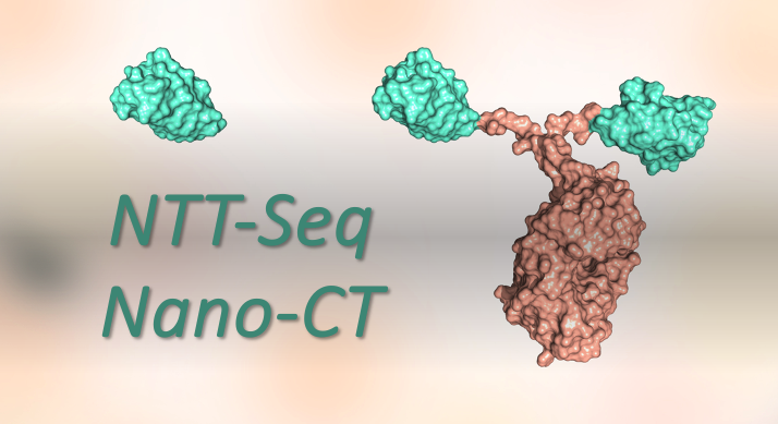
March 11, 2024
The recently developed NTT-Seq (nanobody-tethered transposition followed by sequencing) and nano-CT (nano-CUT&Tag) techniques apply nanobodies - small single polypeptide chain antibodies - to support the profiling of multiple epigenetic modalities at single-cell resolution. Get caught up with the latest in multiplexing and single-cell methods in this insightful blog article!
Read More
<< Back to MOTIFvations Blog Home Page


