THIS PRODUCT IS DISCONTINUED
Fast Activated Cell-based ELISA (FACE™) Kits provide a simple, sensitive method for detecting protein phosphorylation directly in the cell, without making extracts or performing electrophoresis and membrane blotting. These 96-well, high-throughput assays are available in both colorimetric and chemiluminescent formats for over 20 different targets (see list at right). For complete details, click the FACE™ Method tab below.
FACE p38 Kits provide 96 rxns each of 2 antibodies that enable you to monitor and compare the levels of both phosphorylated and total p38. The phospho-p38 antibody recognizes the p38 alpha, beta, gamma and delta isoforms only when phosphorylated at Thr180 and Tyr182. The total-p38 antibody recognizes p38 proteins regardless of their phosphorylation states. Click the p38 Info tab below for data and more information.
| Name | Format | Cat No. | Price | |
|---|---|---|---|---|
| FACE™ p38 | 1 x 96 rxns | 48100 | Discontinued | |
| 5 x 96 rxns | 48600 | Discontinued | ||
| FACE™ p38 Chemi | 1 x 96 rxns | 48200 | Discontinued | |
| 5 x 96 rxns | 48700 | Discontinued | ||
| FACE™ p38 Manual |
| FACE™ Profile |
| Cell Biology Products Brochure |
| IsoCyte™ Application Note – Phospho-Protein Detection |
| MSDS: Sodium Azide |
| MSDS: Sulphuric Acid |
| MSDS: Thimersol |
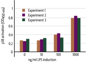
Figure 1: Colorimetric measurement of phosphorylated p38.
Macrophage 4/4 cells were cultured in 96-well plates. Cells were induced with the indicated amount of lipopolysaccharide (LPS) for 20 minutes. Phosphorylation of p38 was assayed in triplicate using the FACE p38 Kit. Cell numbers in each well were normalized using Crystal Violet.
Antibody Specificities
The phospho-p38 antibody is specific for phosphorylated p38 and was raised against a peptide phosphorylated on residues that correspond to the sequence surrounding Thr180 and Tyr182 of human p38. It recognizes p38 alpha, beta, gamma and delta isoforms. The total-p38 antibody recognizes p38 MAPK regardless of its phosphorylation state.
p38 MAPK Overview
p38 MAP kinase is a member of the Mitogen Activated Protein Kinase (MAPK) family of proteins. It was first identified in a screen for drugs inhibiting tumor necrosis factor-mediated inflammatory responses. p38 MAPK is activated in response to a variety of stimuli including growth hormones, ligands for G-protein coupled receptors, inflammatory cytokines and stresses such as osmotic shock and heat shock. Because of its critical role in inflammation regulation and stress response, p38 MAPK is of great interest in both basic and therapeutic research, making high-throughput study methods in demand.
The FACE™ Method
In FACE, cells are cultured in 96-well plates and stimulated to induce the pathway of interest. Following stimulation, the cells are fixed rapidly, which preserves activation-specific protein modifications. Each well is then incubated with a primary antibody specific for the activated protein of interest. Subsequent incubation with secondary HRP-conjugated antibody and developing solution provides a colorimetric or chemiluminescent readout that is quantitative and reproducible (Figure 1). The number of cells in each well can be normalized easily with the provided Crystal Violet solution. FACE Kits also contain primary antibody specific for the native inactive protein, so you can monitor both native and activated protein levels in the same experiment. FACE eliminates cellular extractions, radioactive kinase assays, time-consuming Westerns and inefficient epitope interactions that occur on membranes. FACE is a highly sensitive high-throughput assay designed for detecting activated proteins within mammalian cells.
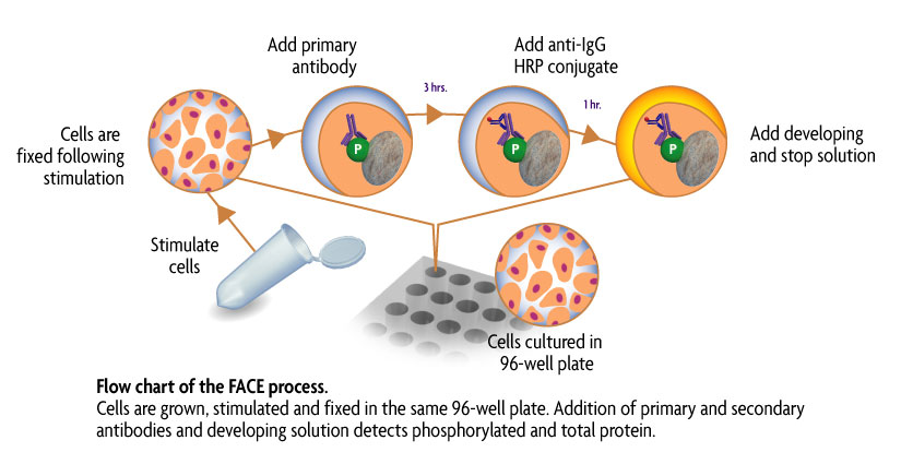
Figure 1: Flow chart of the FACE process.
Flow chart of the FACE in cell Western method that uses a cell based ELISA to measure the levels of the native and phospho forms of signaling proteins and kinases that are activated by phosphorylation.
Contents & Storage
Two (or ten) 96-well plates for culturing cells, 96 (or 5 x 96) rxns each of two primary antibodies (1 phospho-specific, 1 specific for native protein), HRP-conjugated secondary antibody, Quenching Solution, 1X Antibody Blocking Buffer, 1X Antibody Dilution Buffer, 10X PBS, 10% Triton X-100, 1% SDS Solution, Developing and Stop Solutions, and Crystal Violet Cell Quantification Solution. Storage conditions vary from room temperature to -20°C, see manual for details. All reagents are guaranteed stable for 6 months when stored properly.