THIS PRODUCT IS DISCONTINUED
Fast Activated Cell-based ELISA (FACE™) Kits provide a simple, sensitive method for detecting protein phosphorylation directly in the cell, without making extracts or performing electrophoresis and membrane blotting. These 96-well, high-throughput assays are available in both colorimetric and chemiluminescent formats for over 20 different targets (see list at right). For complete details, click the FACE™ Method tab below.
Each FACE ErbB-2 Kit provides 96 rxns each of 2 antibodies that enable you to monitor and compare the levels of both phosphorylated and total ErbB-2. The Y877 kit contains phospho-ErbB-2 antibody that recognizes ErbB-2 only when phosphorylated at Tyr877, while the Y1248 kit provides a phospho-antibody that recognizes ErbB-2 only when phosphorylated at Tyr1248. Each kit contains a total-ErbB-2 antibody that recognizes ErbB-2 regardless of its phosphorylation state. Click the ErbB-2 Info tab below for data and more information.
| Name | Format | Cat No. | Price | |
|---|---|---|---|---|
| FACE™ ErbB-2 (Y877) | 1 x 96 rxns | 48130 | Discontinued | |
| 5 x 96 rxns | 48630 | Discontinued | ||
| FACE™ ErbB-2 (Y877) Chemi | 1 x 96 rxns | 48230 | Discontinued | |
| 5 x 96 rxns | 48730 | Discontinued | ||
| FACE™ ErbB-2 (Y1248) | 1 x 96 rxns | 48105 | Discontinued | |
| 5 x 96 rxns | 48605 | Discontinued | ||
| FACE™ ErbB-2 (Y1248) Chemi | 1 x 96 rxns | 48205 | Discontinued | |
| 5 x 96 rxns | 48705 | Discontinued | ||
| FACE™ ErbB-2 Manual |
| FACE™ Profile |
| Cell Biology Products Brochure |
| IsoCyte™ Application Note – Phospho-Protein Detection |
| MSDS: Sodium Azide |
| MSDS: Sulphuric Acid |
| MSDS: Thimersol |
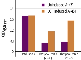
Figure 1: Monitoring total- and phospho-ErbB-2 using FACE.The FACE ErbB-2 (Y1248) and ErbB-2 (Y877) Kits were used to assay the levels of total and phosphorylated ErbB-2 contained within untreated or EGF treated A-431 cells.
Antibody Specificities
The FACE ErbB-2 (Y877) Kit contains a phospho-ErbB-2 antibody that was raised in rabbit against a synthetic phospho-peptide corresponding to residues surrounding phosphorylated Tyr877 of human ErbB-2 and recognizes ErbB-2 only when phosphorylated at Tyr877. The FACE ErbB-2 (Y1248) Kit contains a phospho-ErbB-2 antibody that was raised in rabbit against a synthetic phospho-peptide corresponding to residues surrounding phosphorylated Tyr1248 of human ErbB-2 and recognizes ErbB-2 only when phosphorylated at Tyr1248. The total-ErbB-2 antibody supplied in the FACE ErbB-2 Kits recognizes ErbB-2 protein regardless of its phosphorylation site.
ErbB-2 Overview
The ErbB-2 proto-oncogene, also called Neu, EGFR-2 or HER-2, is a member of the transmembrane receptor tyrosine kinase family, which also includes EGF receptor and EGFR-3 (HER-3 or ErbB-3). Although, ErbB-2 does not have any known high-affinity ligands, its kinase activity can be activated without ligand by either overexpression or heteroassociation with other members of the ErbB family. Amplification of the ErbB-2 gene and overexpression of its product has been detected in almost 40% of primary human breast tumors, which correlates with poor prognosis in node positive breast cancer. ErbB-2 overexpression is also observed in ovarian, gastric, salivary and non-small cell lung carcinomas. Because of this, ErbB-2 is one of the major drug targets for breast cancer and other cancer treatments.
The FACE™ Method
In FACE, cells are cultured in 96-well plates and stimulated to induce the pathway of interest. Following stimulation, the cells are fixed rapidly, which preserves activation-specific protein modifications. Each well is then incubated with a primary antibody specific for the activated protein of interest. Subsequent incubation with secondary HRP-conjugated antibody and developing solution provides a colorimetric or chemiluminescent readout that is quantitative and reproducible (Figure 1). The number of cells in each well can be normalized easily with the provided Crystal Violet solution. FACE Kits also contain primary antibody specific for the native inactive protein, so you can monitor both native and activated protein levels in the same experiment. FACE eliminates cellular extractions, radioactive kinase assays, time-consuming Westerns and inefficient epitope interactions that occur on membranes. FACE is a highly sensitive high-throughput assay designed for detecting activated proteins within mammalian cells.
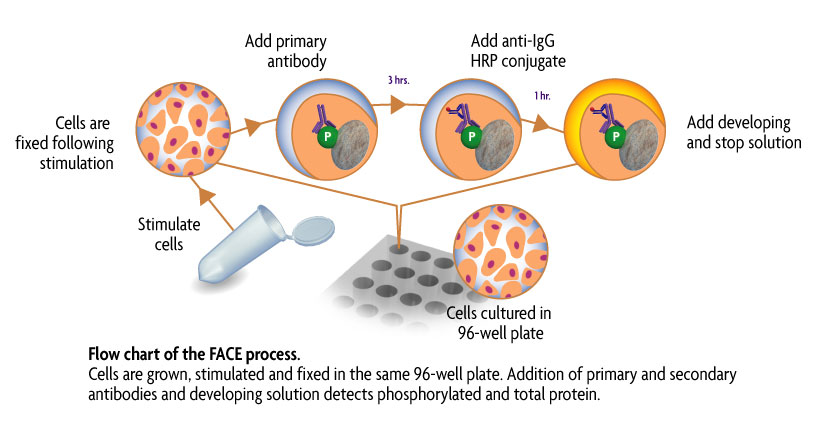
Figure 1: Flow chart of the FACE process.
Flow chart of the FACE in cell Western method that uses a cell based ELISA to measure the levels of the native and phospho forms of signaling proteins and kinases that are activated by phosphorylation.
Contents & Storage
Two (or ten) 96-well plates for culturing cells, 96 (or 5 x 96) rxns each of two primary antibodies (1 phospho-specific, 1 specific for native protein), HRP-conjugated secondary antibody, Quenching Solution, 1X Antibody Blocking Buffer, 1X Antibody Dilution Buffer, 10X PBS, 10% Triton X-100, 1% SDS Solution, Developing and Stop Solutions, and Crystal Violet Cell Quantification Solution. Storage conditions vary from room temperature to -20°C, see manual for details. All reagents are guaranteed stable for 6 months when stored properly.