Recombinant Mononucleosomes H3.3 (R8C) - biotin
| Catalog No: 81287 | Format: 20 µg | ¥4,160 | Add to Cart |
| Catalog No: 81987 | Format: 1 mg | ¥41,200 | Add to Cart |
Request a quote for a bulk order Request Quote
Expressed In: E. coli Protein Species: Human
Contents
A representative Technical Data Sheet (TDS) is provided here. Please refer to the lot-specific TDS you will receive with your order for the lot-specific buffer contents and protein concentration.
Background
In vivo, histones are wrapped around by DNA in chromatin. Therefore, nucleosomes are more physiologically relevant substrates than histones and histone-derived peptides for in vitro studies. More importantly, some histone methyltransferases are signifcantly more active, as well as specifc, when using nucleosomal substrates in HMT assays, such as DOT1L and NSD family enzymes. Nucleosomes are also widely used in histone methyltransferase screening assays to identify small molecular inhibitors for drug discovery. Histone H3.1 and Histone H3.3 are the two main Histone H3 variants found in plants and animals. They are known to be important for gene regulation. Histone H3.1 and H3.3 have been shown to demonstrate unique genomic localization patterns thought to be associated with their specific functions in regulation of gene activity. Specifically, Histone H3.3 primarily colocalizes with marks associated with gene activation (H3K4me3, H2BK120ub1, and RNA pol II occupancy). Deposition of the Histone H3.1 variant into the nucleosome correlates with the canonical DNA synthesis-dependent deposition pathway, whereas Histone H3.3 primarily serves as the replacement Histone H3 variant outside of S-phase, such as during gene transcription. Histones are linked to tumorigenesis primarily through alterations in their PTMs and the enzymes regulating these modifications, suggesting that they might disrupt the reading, writing, and/or erasing of these marks. Except for being near K9, which can be methylated or acetylated, R8 site itself can be methylated too. Mutations in histone H3 occur with high genetic penetrance within rare paediatric gliomas and sarcomas. Researchers found that mutations at H3 N-terminal residues at or near PTM sites including R2, R8, K18 and R26 might act as oncohistones.
Application Notes
Recombinant Mononucleosomes H3.3 (R8C) - biotin are suitable for use as substrate for histone modification enzymes, or to generate chromatin in vitro.
Protein Details
Recombinant Mononucleosomes H3.3 (R8C) - biotin consist of a 167 bp of 601 DNA and two molecules each of histones H2A that includes amino acids 1-130 (end) (accession number NM_003512), H2B that includes amino acids 1-126 (end) (accession number NM_003518), H3.3 that includes amino acids 1-136 (end) (accession number NP_002098.1) with a point mutation Arg8Cys, and H4 that includes amino acids 1-103 (end) (accession number NM_003548). All of these histones were expressed in E. coli cells. The molecular weight of histone octamer is 108 kDa.
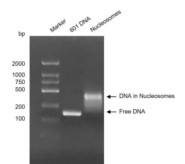
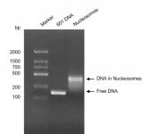
Recombinant Mononucleosomes H3.3 (R8C) - biotin, DNA gel
Recombinant Mononucleosomes H3.3 (R8C), biotin, were run on a 2 agarose gel and stained with ethidium bromide. Lane 1: DNA marker. Lane 2: Free 601 DNA which was used for assembly of nucleosome. Lane 3: Intact mononucleosomes H3.3 (R8C), biotin. Intact mononucleosomes H3.3 (R8C) migrated much higher than free 601 DNA. The agarose gel shows that almost all of 601 DNA wrapped histone octamers to form nucleosomes.
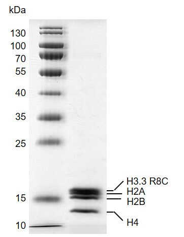
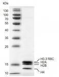
Recombinant Mononucleosomes H3.3 (R8C) - biotin, protein gel
12.5% SDS-PAGE gel with Coomassie blue staining
MW: 108 kDa
Purity: >92%
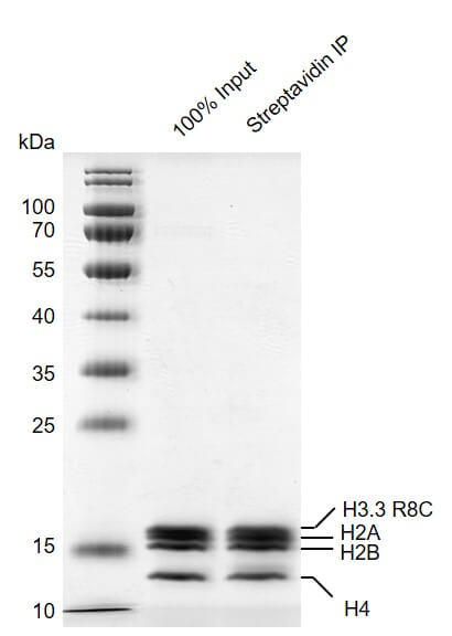
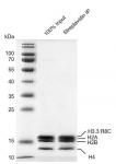
Streptavidin pull down assay for Recombinant Mononucleosomes H3.3 (R8C) - biotin
24 µg biotinylated mononucleosomes were incubated with 10 µl streptavidin beads for 1 hr at 4°C. Streptavidin beads were washed 3 times with 1 ml binding buffer. Then the beads were added 60 µl 2×SDS loading buffer and boiled for 10 min at 95°C. 2.4 µl samples were loaded and run on a 12.5% SDS-PAGE gel and stained by Commassie blue. The SDS-PAGE gel result showed that almost all of biotinylated mononucleosomes were pulled down by streptavidin beads.
Storage
Recombinant proteins in solution are temperature sensitive and must be stored at -80°C to prevent degradation. Avoid repeated freeze/thaw cycles and keep on ice when not in storage.
Guarantee
This product is guaranteed for 6 months from date of arrival.